
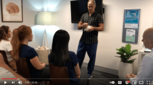
Guest Speaker: Dr. Marc Coghlan
Facilitator: Matthew Hodgson
Participants: Chiropractors, Physiotherapists and Medical Doctors at Platinum Chiropractic Erina
Dr Marc Coghlan Neurosurgeon: “Different groups and particularly chiropractic group I have got a lot of interaction with Chiros. And quite often we’ll chat about patients. And I really think that’s a good way to practice. And, you know, I kind of, I wish a lot of my other colleagues will also embrace a more holistic approach. We don’t have all the answers. As a group, as surgeons, we certainly don’t. And I’m sure you guys feel the same. So, I think, collectively, we can probably practice better for the patients in terms of putting our knowledge together with Chiros, Physios, GPs rather than kind of all working maybe separately here with the same patients. And the other thing is, I mean, I guess the role of surgery for most patients that you’d see is obviously minimal. So, you know, I might see, oh gosh, if I see 30 patients in a day, you might find a third of them, or not even, sometimes even a quarter that actually need a procedure, or sometimes even less, and those are patients that have been seen by GPs, and then referred thinking as surgical candidates.
So, the reality is, don’t take this talk out of perspective, the reality is, most patients actually don’t really need surgery. And the ones that we sort of operate on, we really have responsibility to picking out those patients that we think are going to do well with surgery. And that’s a big skill. And that takes a lot of experience. And I know, some good surgeons, and then I know some bad surgeons technically, but then also the other whole side of the discipline is choosing who to operate or who should we operate on. And sometimes that’s quite hard, because, you know, you think patients might do well, and some do poorly and you really have to try and I guess, tease out those that are good surgical candidates.
The other thing that’s crucial is whenever I see a patient, I really, you also have to weigh up the natural history of the condition and then look at whether you operate or not. I mean, there’s no point in intervening if the patient’s coping and you know, they’re actually going to get better. I mean, you got to kind of practice, I think you got to practice quite ethically and honestly and say to the patient, these are your options, you know. So, also at the end of the day, it’s important, very important to be honest with patients, and we say, this is a natural history and this is what we can offer. And so, we shouldn’t really be in the game of servicing people and operating, you have to really tease out who you can help.
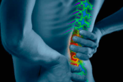
And that’s really important looking at natural history. And we all know like people with disc herniations, a lot of them get better. But we see a lot of patients that have so much pain that they don’t want to wait. I just saw guy now before I left, he just broke, he was in tears, he hasn’t slept for a month. Severe Cervical Radiculopathy, literally suicidal. So, there’s no point in saying go for four months on Lyrica. There’s also a lot of literature coming out on the adverse effects on drugs, you know, Lyrica, I think personally, I personally think in this country that we’re going to look back one day and say, we shouldn’t have been prescribing this whole plethora of opioids. I think Australia is in a bad way in terms of, you’ve got pain, reach out and get an Oxycontin or something, I think it’s absolute nonsense. And I think unfortunately, that’s something the medical industry has kind of inherited and passed on from maybe previous doctors, you know, and that’s something that has to be changed as well. Can’t just treat pain with opioids because you end up creating much bigger problems. And you end up creating two problems, dependence and pain, it doesn’t fix the pain.
And but, you know, neurosurgery in particular is a really, really exciting field. Most days, we probably spent about half our time using the microscope, doing sort of microscopic surgery. And then the other half doing more macroscopic surgery with the loops, where it has got maybe a bit more of an orthopaedic flare. I remember working as a registrar and doing some of the old-fashioned fusions and just absolutely hating it. Opening everything, muscles bleeding, you’ve got the nerves exposed, you’re attracting the nerves and you are trying to get to the disk space. And then, you know, putting in screws and rods and closing up anything shit, like this. There’s no way I want this done in my spine. It’s just like barbaric. And that’s how we were trained. That was like the gold standard like to.
And then with new technology, it was fantastic coming in Australia, we had great opportunities where we could go to Japan or we go to America and all these courses. And we started learning, being exposed to different techniques. And some of the techniques that I’ve adopted, that I’m really passionate about, particularly approaching the spine from the front. And then also the last 10 years doing a lot of lateral approaches where we come in from the side. And the big advantages of some of these approaches or if you can do an operation without cutting tissue or violating tissue and you just separate tissue, it’s so much less traumatic and the patients really feel the difference.
So, a classic example is operating on the neck like if you use this, like a natural cleavage plate, and we literally, you can use like a cotton wool band, and you can separate those muscles. And if you look at your lumbar spine, between your two big layers of Longissimus and Multifidus, there’s an interfacial plane. And you can see it on the MRI, and that’s called a Wiltse approach. And it’s, actually perfectly kind of engineered, if you look at that there’s a muscle bulk there, and a muscle bulk there. And then there’s interfacial line, and there’s no blood vessels there. And sometimes when we do it, it is called a Wiltse approach, the guy was a surgeon from the States, you can do a small little incision, and I literally just use my finger and I run my finger down there and it separates those two planes. And that takes you right to the edge of the transverse process. So sometimes these days, if we have to put like pedicle screws in, that’s how we do it. And so rather than stripping on the muscle.
And a tell-tale sign of it is if you look at the post-op MRI’s, sometimes you can see this horrendous tract of scar tissue. And if you know you’ve done a good job, if you look at it and you don’t see any scar tissue, muscles folded back. And so those are kind of some of the advances. So, that’s one of the techniques that we do try and employ.
But I think let me go back a step and say, there’s also an old adage, you probably all have heard, “keep it simple and keep it safe”. So, patients that do really well are patients with like severe nerve pain, where there’s a predictable surgical target, you go and take the disc up, free up the nerve, they just tend to be really happy and almost on the ward round on the next day, they say my nerve pain is gone. And you know it’s going to be. And so traditionally, it’s very important that we would try and be careful operating on axial, non-radicular back pain. In other words, someone comes in with back pain, be very careful. And you can imagine why, that’s an absolute quandary as to what the pain generator is. Imagine all the different pain generators, the muscles, discs, sacroiliac joints, facets, but that is also a little bit antiquated. And I think we have to be more proactive.
So, these days, we use bone scans, we use diagnostic blocks, MRIs, and you can try and narrow it down and work out on what’s causing the pain. But you know so and quite often we’ll sort of even do a discogram, we’ll get a radiologist to check one disk, and to see if it’s discogenic pain. So, the crucial thing is that every patient has to be meticulously worked up so that by the time you’re operating, on them, you know, is it discogenic pain, is a facet pain or is it sort of SI joint pain and various other entities that won’t respond to surgery. And the reality is, as you know, for your patients, often it’s a mixed bag. You know, so we have to be a bit cautious with discogenic, or sorry, with, disc axial non-radicular back pain.
The other point I want to make, Matt and I were talking about a case. Sometimes people think nerve pain is nerve pain. But you know, if you look, this is a 3D printed model of someone’s spine. If you look at this, this is the reality of what we’re dealing with these as a lot of older people, and they get a lot of spurs, it’s all biomechanics, as bone rubs on bone, you get these massive platforms that form. And we see a lot of patients also with like foraminal stenosis, and you can see how, if you look at the foramen, and when you see patients, you should always try and work out, is there nerve compression in the canal, or in the foramen, because it’s completely different in terms of what it’s going to respond to. If it’s in the foramen also look at it and say, is that foramen enclosed in a vertical dimension like that, with a pedicles sort of sunk down? Or is it in an AP direction due to large osteophyte, which this patient has got as well digging back in.
So, what we found is when the patient is laying recovered like that, if you decompress the canal, and it’s pretty easy, they tend to do well. If you go and drill out the foramen, it doesn’t really help them because once they’re on their feet, they’ve still got that vertical closure on the foramen. And that’s why some of those patients need like an interbody graft, we actually put a spacer in. And so, the problem with doing sometimes small operations, and if there’s a high rate of recurrence where they don’t work, because of them collapsing, you just end up creating a lot of scar tissue. So, as we get more experienced in putting like spaces in anteriorly, quite often, even people in their 80s and 90s, if they’re really struggling, we’ll put a spacer in. And that’s one of the sort of things that I’ve adopted over the years.
I just want to show you. I brought this model. That’s like a sort of traditional anterior spacer with a plate but what we found was a lot of these spacers, load in various points because this is each person’s geometry of their body, so different. And of late the last couple of years, it’s been a really quite an exciting journey. We’ve been sending scans to the engineers and they make these 3D reconstructions. And then they give us various like 3D printed spacers to fit. I think this is the wrong one. They actually fit, they actually fit perfectly in, so you can see that. So, it’s quite interesting. We did some studies on Cadaveric bone, like if you load that, compared to a normal spacer, because it’s a perfect surface match. It is so much stronger, you don’t get point loading where the sharp edge of the cage goes into the edge of the bone. Imagine stepping onto sand, and you’ve got this perfect match with the sand, it’s just the distribution of forces is so much better.
So, this is something that I’m really passionate about. What we find is we’ve seen quite a few old people with like Scoliosis, where previously we wouldn’t have taken them on. And now we are sending to the engineers do a scan, and they do a recon. And they say to us, to get that patient back in alignment, these and say you’re doing like a four-level anterior spacer. These are the different spaces you need. And then it’s quite good, they give us like a virtual surgical planning. So, there’s some pre-surgery and they give us some model post-surgery. And that’s where the concept of sagittal balance is crucial. And I think that’s where there’s a lot of overlap with Chiropractic, and surgeons.
In the past surgeons do nothing about sagittal balance. They used to put rods in, imagine putting a flat Harrington rod in a Lordotic spine, it just doesn’t work. And then in a couple of years, the next level is completely shot. What’s really, really interesting, if you fuse someone’s spine, in good lordosis, they’ve shown studies to show that the next level hardly deteriorates. So, we always thought if you fuse it, they’re working harder, therefore it’s going to fail. But it was the way it was fused. So interestingly, if you actually fuse someone in a nice perfect Lordosis, and the next disk is parallel and not sort of working harder, it’s incredible how much longer it lasts. So that’s the kind of thing where maybe people think, okay, if you fuse it, the next level is going to fail, it’s not as how it’s done. So, if you fuse it so that is in a neutral, sagittal balance. Sagittal balance is a concept of when you’re standing side on, your C7 should be vertically above your sacrum.
And if you see a lot of our elderly patients, they are becoming more kyphosed, as they become more kyphosed, they’re putting a lot of torque on their lower back. And if you do an operation that makes it worse, they are never good. Because it’s the centre. And you’ve probably seen them, they all do this, they start rotating their pelvis and bending the knees to compensate, and then they start doing this. This is terrible!
Yeah, so those are the sort of parameters that we sort of look at with each patient before you take them on. And the technology, this technology has allowed us to, Matt and I was saying, you know, we were talking about would you do four or five levels, we do, especially the older people now and it’s allowed us to take it on. But you’ve got to be cautious.
And the other thing is, while we take disc replacement technology, the funny thing about disc replacement is, everyone, there’s a lot of misinformation. A lot of patients want disc replacements, and you’ve also got to be cautious to explain to them that you’ve got to match the pathology, the technology with the pathology. So in other words not everyone is suitable for disc replacements. And this is I’ll just pass this around. This is an American disk that I’ve been using called active L. And it gives you an idea. It’s basically, it’s a polyethylene core on a very hard metal alloy that doesn’t have any like particular debris or wear and it articulates and it’s quite clever. It allows flexion and flexion extension, lateral bending, and it even allows a little bit of translation. So, this little core translates within about two millemetres.
But the newer discs are going, this is still, think about it. This is still a ball and socket type model. The problem with that is you put that into a disk and then you’re now forcing your facets to move in this sort of predetermined arc of rotation. Our natural discs are more like an elastic type thing. So that’s why we also see quite a lot of facet driven pain with disc replacements; I believe anyway.
And to give you an example, you got to be very careful of putting a disc replacement in a steep environment. Think of patients with their par’s fractures, L5 is wanting to slide over the sacrum. And it creates a lot of stress fractures or stress forces through that area. So, those patients realistically actually do better with a more stable solid like a 3D printed-spacer. If you put this into their environment they don’t do so well. I’ll just pass this around. It’s worth just having a look at it in terms of the biomechanics.
Matt Hodgson: “Marc, can you just go through, what you’re looking at when you’re choosing between a disc replacement and fusion? Why you would go one to the other?”
So firstly, if you’re going to do a disc replacement, they’ve got to have healthy facets. Imagine if you’ve got severe facet joint where you can’t go and put in a mobile disc, it would make those facets articulate too much, and those would cause a lot of facet pain. So, we grade facets, generally grade one to four on CT. Grade one with a very mild problem, you can generally get away with the disc replacement. But if you’ve got really bad facets, there’s no point in putting it in. I mean, your facets have to move synchronously with your disk. And if these are bad and locked down, it doesn’t make sense.
The other thing is the level L4, L5, I’ve noticed the disk replacements at four, five, tend to do much better because it’s a nice horizontal disc. So, you don’t have that translational or sliding force. And in what is quite a good concept is a concept of a hybrid, where, if they’ve got two levels of discopathy, bad discs, you put in a stable one and you actually put in a very low dotty cage to bring L4 end plate horizontally. And then you can do a disc replacement at L4. So, we try and mix and match things, according to the patient’s pathology. But you do have to be careful because you can end up causing facet pain. And it’s not always plain sailing.
Another thing we haven’t spoken about, I think it is extremely important. We’re talking about natural history just to digress. Whenever you see patients with cervical issues, always take a few minutes to test their reflexes. Because a number of times I’ve like, picked up patients that are myelopathic where they’ve got cord damage just from doing a simple reflex. And it’s a reminder, especially for me, when I’m busy, and I’m just looking at the imaging. But the number of times I’ve sort of picked up things just from doing a basic reflex, and they tend to be, they’re hyper reflexive, they’ve got early spasticity, it gives you a clue. And those patients be very careful with any sort of exercise, anything you put them through. For me, that’s a one time I’d be very careful in terms of hyperextension, especially in the neck.
And if the MRI scan shows a good buffering layer of CSF, you’re generally safe. And if it shows that there’s a really cord compression with something called Myelomalacia, those cases are very, very dangerous. I mean, we find even if we position them in hyperextension, they can deteriorate because think of the ligamentum flavum and the discs in a dynamic phase. And on the MRI, they could be okay. And in a certain position they become hyper extended, they can start damaging their cords.
So always, you know, try and have a sort of, don’t just rely on imaging and then try and really also have a clinical knowledge, spend a bit of time clinically examining the patient.
Matt Hodgson: So, how does your approach differ with someone they have, that has myelomalacia compared to just DJD? And then how do you counsel people around that?
So, if they’ve got established spinal cord damage, generally we would use it as an indication for surgery. Most surgeons say once would agree, then once you’ve got that, it’s called a high signal intensity changes on MRIs. And the best MRI to look at is the one with a spinal fluid is white, T2. And you’ll see whiteness or edema within the cord. And those patients generally need surgery unless they’re really elderly, and there’s other confounding factors. But the problem is we do counsel them and saying that the natural history of that, even with surgery, two thirds chance that you’ll improve, and one third chance that you might just stabilise. But most of them get better. But there’s a small group of patients where they’ve got really bad fibrosis, and they end up with a lot of burning in their feet, it is quite frustrating. And you do a wide decompression and it doesn’t change, but they are small group. But that’s crucial picking up those patients because you want to stop that natural deterioration with Myelopathy.
Neurosurgeon Dr Marc Coghlan
Matt Hodgson: So, if you’re referring those people on to physio or chiropractor, that sort of thing, how prescriptive are you in terms of what you think we should do?
Yeah, I would be very strong and say don’t do any hyperextension because it’s, we’re using a lot of dynamic MRIs, especially the MRI in real time where we assess a patient for flexion and extension. It is fascinating when they’ve got tight cords to see the difference when they’re hyperextended, it is like really shuts down and then a neutral phase and little bit of flexion opens them up. But in hyper extension it’s you can’t believe the difference. So that’s another thing is, you know, if you look, don’t think of the spine, just from the imaging, think of it in a dynamic phase. And we should really be utilising like standing x-rays, or standing upright MRIs. Because, I mean, you know, we’re not lying down all day.
Matt Hodgson; So, what do you, for you if we’re referring to you what would constitute a good referral and what would be an emergency referral, where we should say, what would be the signs?
So, probably the commonest emergency referrals would be patients who have got sort of unremitting pain and associated with some sort of neurological differences. A common one we see is foot drop. It is quite frustrating. Sometimes we see foot drop has been sort of treated conservatively for a long time. And that’s foot drop is a really small thing that can be devastating thing for the patient. As you know, it can be devastating on stability, especially the elderly. They’re a nightmare with falls and devastating on their gaits.
So, I honestly think anyone with foot drop, be careful to sort of persevere with treatment. Because it’s, I think it’s a bit of a double edge sword. If you keep them too long and don’t refer them and then don’t get better, you’re better off referring them really early and saying you might not have to have surgery but go and be assessed, have a nerve conduction test done. So, foot drop, definitely. My absolute nightmare is old ladies with bladder dysfunction. It just makes me run a mile because, you know, you don’t know. And sometimes they actually get worse with surgery, especially if they’ve got instability, they’ve got pelvic floor problems, they’ve had a lot of children. That’s a really difficult thing to assess. But the classic corda equina where people have bladder and bowel issues, be very careful with that. Because those just do not recover.
And then as I said, patients that are hyper reflexive where you examine and, there’s very brisk reflexes, something’s compressing on their cord, I would refer them early. And patients with cord signal change. So probably those foot drop, bladder and bowel, cord signal change. Atrophy as well. But unfortunately, often, you know, by the time you see someone have a lot of atrophy, that some of that is sustained already. Alright. But your bladder, bladder and bowel, that’s also something I guess you just have to check with a lot of patients with, sometimes they don’t want to talk about, just ask them. Because it’s easy to sort of just glimpse over.
Matt Hodgson: So, if when we refer someone to you, how soon after surgery, would you be saying to the patient, you know you need to go back to your chiropractor or your physio to manage the other level?
Yeah. So, I think it depends on the pathology, it depends on the patient. I think some of our patients, especially some of the elderly, there is a group and I really just say to them you should just sort of rest for six, eight weeks, just like recover. And I’m always scared of sending some of those patients back to physios or chiros in the community, if I’m not sure what they’re going to do. Because some of them are osteopenic. You put these constructs in and the last thing I wanted someone getting them to like do something that they haven’t done for 10 years. So, I just told them just eight weeks and but then the basic, the basic stuff, you know, where we’ve done like the decompressions and that really soon you know, get them moving, probably within about four weeks, three to four weeks after surgery. Yeah, because the key there is movement, being active, and not just kind of waiting for things to settle down.
Does anyone else have any questions?
Are you doing the disc decompressions in the neck as well? You’re only talking about lumbar spine.
Yeah, we do. We do a lot of them in the neck. And when you look at neck surgery, there is two big categories of surgery. There’s one where you do foraminotomy. So, the neck anatomy is completely different. So, foraminotomy is if you get like a soft disc and it’s burst and it’s pushing on the nerve, it’s a really good operation where we come in from the back, we flex the patient a little bit on the table, and we use that interlaminar window and you can see that’s a facet joint, we just drill a tiny little window medial to the facet joint, expose the nerve roots and generally you can sort of tweeze out a little bit of disk. So that’s good if they’ve got arm pain.
And then a lot of elderly patients don’t have the soft discs, they’ve got the foraminal stenosis. It’s also very nice common operation, we just widen the foramen, we use a little two mould drill with irrigation. It’s called egg shelling. We just drill until you can see the thecal sac. Remember this is a cord but the reality is you’ve got a nice layer of protective dura and then you’ve got the ligamentum flavum. So, I actually drill and I use that ligamentum flavum as a little bit of a buffer. And you draw until you can see through the bones like an eggshell, and then you take a hook, and you just flake that off. So it’s really quite a nice operation.
And then the other big category of surgeries, is when you come in from the front, very common. That’s really good if people have neck pain. Axial neck pain and especially, you know, a lot of our patients have kyphosis. And you won’t believe it, every single week, especially ladies, I see ladies that come and say, Doc I feel like my head is too heavy, just these headaches, occipital neuralgia, and I feel like my head is too heavy for my neck. They do terrible if you go into a laminectomy because you’re releasing that posterior tension band, and you’re worsening their kyphosis.
So that’s why a lot of the old surgeries I think, made people worse in the long run. You got to think very biomechanically, before you choose what surgical approach. Another point if someone who’s kyphosed and doing a laminectomy, I mean, I see these horrendous CTs with all the bones being drilled out. And the patient is you know coming in like this, obviously, you can’t release. This is interspinous ligament and that acts as a posterior tension band. But once you release that, especially in the elderly, they’re going to become more kyphosed. So, if someone comes in with a beautiful lordosis, and you need to decompress them, you have that latitude to do that. But if they come in, and they’re really kyphosed, you can’t go into them. So, yeah so keyholes, from the front and cervical disc replacement, that’s also a really good operation.
And I have to say one thing, the neck is much more forgiving than the back, to operate on. Because think of the different biomechanics. This takes so much little less weight than the lumbar spine. So, we always joke at conference and say, you can get away with a lot more in the neck in terms of patients recovering quickly.
And how do you choose whether you’re doing an anterior or posterior approach?
Yeah. It is anatomy. So, I mean, I’m sure it’s the same with anything, you can’t do a good job if you don’t know the anatomy. So, you look at the imaging. If it’s a tiny posterolateral disc, it’s pushing on the nerve and it’s away from the thecal sac and the cord, you can sneak in from the back and take it out. But think of a lot of the discs that you see on MRI, they start almost centrally, and then like a broad-based thing that goes up into the foramen. There’s no point in tweezing out a tiny bit of that and causing fibrosis, and you leave a whole chunk of ruptured disc anterior, you know.
So, we look at the anatomy. And generally, you’re going to get a much better clearance from the front, because you can see from side to side. Do any of you pay attention to this little, probably the most neglected joint in the spine on the corners? The uncinate joint. And it is amazing how many people like have neck pain and you do a bone scan, and the disc has deteriorated and they get this sort of sclerotic bone on bone loading with like, ivory bone in those, and we call them uncovertebral joints. So that’s just something to think. And that’s why they are so happy if you put a spacer in. I think we just by jacking it open, yeah it just takes the pressure off and it’s a really annoying pain that exists. That constant neck pain that doesn’t get relief with cortisone injections.
What I’m going to ask you guys, because for years, I’ve been telling people about inclinometers and traction, what’s the evidence that Ligament traction helps? And how long does it help for? Does it only help while you’re hanging upside down? What are your thoughts on it? Because I often tell patients go and lay on an inclinometer, 15, 20 minutes a day, as long as you’re healthy, you don’t have high blood pressure, and it’s going to distract that foramen, maybe transiently reduce the nerve oedema.
Obviously, when you’re back on your feet, it’s negated. But I have seen it help patients. And I’ve also seen it help patients, where you have big ligamentous disk. And there’s a big strong ligament called the PLL, that we see when you operate. It looks like an elastic band. It runs along the back of the spine. And if you see what happens when you traction patients, that PLL’s tension, and it does sort of approximate things. So, do any of you, I don’t know if I’m kidding myself. But I do think it might be valuable? What are your thoughts? Be honest on that.
It does vary from patient to patient, and it depends on their stability too. Because I don’t like the ones, where they’re already spondylolisthesis. But I think with a lot of people that are mostly doing it repetitively, if they can commit to a routine, but I usually always start very small, short amounts of time. And they will vary from patient to patient, but they can generally progress and find longer, weight bearing relief after they’ve done some traction besides early morning or if maybe they’ve warmed up and they walk.
And when, as a chiropractic group when you train traction techniques, do those techniques keep your head in a neutral position or do they cause extensional flexion? In reality, because that’s something I often think about, I know the physios use various things to pull on or inflate collars but I often wonder if it’s pure distraction with extensional flexion. Difficult to know.
We only learnt flexion distraction. We didn’t learn any other function.
So, flexion and distraction?
And everything’s kept neutral on the table. The table actually drops and you can create different axes.
Yes.
But and I think the patient is always neutral. And there’s nothing happening out.
What’s the worst nightmare for chiro in terms of like the patients you don’t want to see?
The reason I’m asking is like, for me one of the worst operations I do and I do quite often is elderly patients with like C12 severe arthritis where they’ve tried everything and that whole joints completely gone. And we do a procedure using the body tom, the inter-operative CT where we put in a screw in C1 and a screw in C2. But you know, they are so careful of the vertebral artery because we’ve been putting these sharp screws basically above and below. So, we do a CT angiogram before, but it’s just one of those procedures. As I’m getting older, I hate doing it because you kind of hard to you know, it is just really, it’s quite nerve wracking until they wake up. And, but intraoperative imaging makes it so much safer, because you can see on each plane, but still, having said that I just, you know, just my risk appetite gets less as I am getting older. And I was wondering if there’s any sort of patient that walks through the door, you guys think you just can’t wait, you know, whatever you do. I’m sure each profession has them.
A slight side track, in vertebroplasty and so, we so we’ll see a crush vertebra. You can see a lot of thoracic wedging. Where do you stand on vertebroplasty?
So, Vertebroplasty, I think, in the right patients is a fantastic, fantastic procedure. You know a Geriatrician sent me a text message other day. But the government withdrew the rebate for it a few years ago. It was really like it was really I think, based on, I thought it was prematurely done. The New England Journal of Medicine study that was done saying, there was very little benefit but like for a lot of these studies, if you analyse them and you look at who’s driving them, it was quite flawed.
So, for me if the fractures are quite acute, especially if they are involving, particularly like the superior endplate for some reason, the elderly soft bone, they do amazingly well. But if you see, like I saw a guy yesterday, young guy, osteoporotic, he has been on opioids, he’s got multiple levels of wedging, it wouldn’t make biomechanical sense to go and inject a hollow cement in each level because there’s not one particular pain generator. He has kyphosis, he’s got like 10% of wedging of his whole thoracic vertebrae. And I’m not going to fix, you don’t fix your posture with.
If there are sort of there were one or two segments and it was more traumatic. Would it be more applicable?
Yeah, they do well. But then again, if someone’s young, I saw a few like snowboarders you know, in a lot of pain. But you said look, you’re actually going to get better at six weeks. So, don’t bother with it. But the elderly patients do amazing with it. But you’ve got to be careful, if you inject cement in, I’ve seen quite a few patients with the next level in fractures, because now when they’re moving, we’ve got this incredibly hard structure and it’s amazing how it just has a domino effect. Yeah, but definitely a good procedure. We should be allowed to do, it’s frustrating.
And another thing is like we do have a lot of patients that we see that say, “Doc, I’ve got this leg pain, I don’t have a fusion what other technologies do we have in?” Years ago, we used to have these fantastic little devices called the interspinous spacer devices. And what they do is and it really would implore you. When you look at patients scans trying to work out if this is foraminal stenosis and try and break it down. So, a lot of those patients when they stand, there is a thing called a superior articular syndrome, where the superior articular facet of the one below, rides up into the nerve root. And that’s the dorsal root ganglion that comes out. The dorsal root ganglion, that’s a part of the nerve that’s exquisitely sensitive. So, that’s what Lyrica works on.
When you see patients that are in agony, it’s not often it’s not the ones in the canal, it’s the ones in the foramen. It’s like a little bulbous body on the nerve and that has a sensory supply. And as they stand, in particular, the elderly, and they get a little bit of facet joint wear and it becomes, and it does that. That hard, unyielding bone drives up and it pushes it into the pedicle of the foramen. And those patients like that’s the type of thing where we used to just put in these little devices, and they were really good. But then the TGA, had a lot of old antiquated surgeons that didn’t know much about that and said no we don’t have enough technology. We don’t have enough randomised trials and they like pulled up like, but those are good technologies that I’m hopeful might come back. But they were unfortunately, they were introduced by companies probably with not enough studies. You know.
So, what’s an alternative? Do you actually shave off the superior part of the inferior facet joint?
A lot of patients that you end up decompressing, you can end up doing hydrogenic damage. Think about it. If someone comes in and they’ve got a party scoliotic segment, partially subluxed damaged facet joint, the horse has kind of already bolted. You then go and start drilling out their facet to decompress the nerve. You make it worse. Exactly!
So, the beautiful thing about those devices, it allowed us to indirectly distract it without weakening the facets. And you always have to say to patient, I always say to patients, when we’re doing a micro disc, we’re not repairing your back, your disk is worn out and the other discs look okay, so it’s already a weak spot, it’s then burst eccentrically on one side, we’ve taken out that piece, I’m not putting in a new, you know, we haven’t put in a disc repair kit or anything. So that’s a reality. It’s not. And that’s why you get things like recurrences. And that’s why they go into post discectomy syndrome, they have back pain.
Has anyone else got any questions?
Yeah, I have a patient with a lot of anterior endplate damage. Do you see that affecting the descending aorta or other organs anteriorly? Because we talked a lot about posterior stuff but.
Yeah. And you know because they’re so slow, they tend to just deflect them, push them out the way. But we do sometimes see some bizarre like syndromes where they compress a vein or compress an artery, but not commonly. We see more problems when we operate on them. And we, try to swing it, very easy to swing their aorta in the common iliac vessels, which we do for every anterior case, on nice anatomy like this.
But that’s a problem we encounter. And that’s when we get quiet bleeding when you’re moving the vessels, when you’re trying to get it off an osteophyte to expose it. So, that’s when it causes problems.
And of course, you know, we do see this particularly in some older patients at the back it’s called OPLL- Ossification of Posterior Longitudinal Ligament, and that’s really hard to deal with because it actually sticks to the dura. And we see that in the neck. And we actually see it a lot. It seems to be like a genetic preponderance to that OPLL. But the one thing, I don’t know how much time you guys have to really get into neuro anatomy, but take the time to spend as much time as you can looking at anatomy because A- it is going to make your work more enjoyable. And B- you’re gonna have a little more confidence what you’re dealing with, you know.
I just have a feeling that a lot of the trainees today, maybe their neuroanatomy is not as good as you see, in the old days, all the physios and everyone used to go to, I remember in South Africa, they used to join us in the anatomy dissections for you for two or three years, you know, and you really have to know your anatomy and spend a bit more time than you would, otherwise going back and really understanding the anatomy, because you’ll actually, it just makes your work more enjoyable, I think.
I had a patient just last week who actually referred to you through their GP so you’ll probably see them soon with canal stenosis and foraminal stenosis and chronic back pain. Would you, I’ll probably give you a buzz or something once she has seen you. But is it the kind of thing where you would go in you and if there’s multifaceted issues because you don’t know the source of the pain would you do multiple things in one hit or are you just going to do it disc or facet or?
No, you are better off. I think you’re much better off, you’re much better off just keeping it simple. So, someone, every day we actually see some severe stenosis and terrible sort of discs and you say them, “What worries you more, your legs or your back?” And if you can get away with just doing a decompression, just we are trying to, you are better off practising simply and conservatively. Because think about it, the more prosthesis you put in and, the more you alter anatomy and the more you alter the movement on the physiology, the more potential for problems. So generally, I would always say, look, we can do this. You might have to come back for part two, but I’d rather be kind of under service you than go nuts and do too much.
That is pretty much what I told her. Exactly! Because I said you’d probably do that.
Because, also remember we’re not treating scans. I mean, we all know, you see some patients who say, “Oh, my God, you must have terrible pain”. But they said, “No, my back doesn’t hurt, it is just my neck”. You say “You’re kidding! Look at your spine. It is dreadful”. I mean, you see it every day. So, correlation between imaging, and pain is like so poor. That’s what turns the whole science upside down. So, you know, I think if surgeons are saying to patients, “No, look, this looks terrible in your scan, we have got to operate it. That’s fraud science, think about it.
And then, the real challenge is, well think about patients with severe stenosis, okay. They will present with mostly presented with leg pain. But there are a cohort of patients where they get axial back pain, or buttock like ischemic pain. And those are a little bit difficult to tease out because we do see a lot of patients where you can say we’ll do a decompression, but might not actually help your axial back pain.
Matthew Hodgson: So, around that, do you have any sort of examination pearls, that you use to differentiate these symptoms?
Yeah, I mean, I think, so reflexes are always good. Just check the reflexes. But reflexes aren’t that helpful in elderly with general stenosis. Straight leg raise, I always do it, but not always that helpful. Always look at tibialis anterior, so always get them to do that movement. That’s probably the most important movement, you should check every time. Extensor hallucis longus and tibialis anterior.
And the other thing is, I mean, you’re actually going to get the most knowledge just watching the patient walking into your room. By the time the patient sat down, you should already be saying it’s hip, trochanteric bursa or it is neuropathic. So, you see the classic, we will see the straightaway and I say to them, shit they have come to the wrong place. And, then you get a mixed bag, and the worst of those with nerve compression, trochanteric bursitis, and pain down the ITB, you sort of. But don’t operate on them, send them for an injection. If, they get better, tell them they can come back if there’s stenosis present. But I mean, the number of people that can treat you for sciatica and they’ve got-
And for some reason I can’t explain it seems to go hand in hand with people when they get nerve pain, they’re in bed, or they are less active and then they burst the trochanteric bursa. It is really bizarre. It’s almost like someone’s throwing a curveball to confuse us. But, it’s common. It is really common.
In terms of like examinations, with you looking at cervical myelopathy, what are your tests?
So, I always look at it first, are they hyper- reflexic or areflexic, straightaway. Upper motor neuron, cord myelopathy or a nerve issue. Strength, grip strength, especially in the neck. Atrophy. Always look at the muscle groups for symmetry, especially C5, is a really bizarre. C5 is the most sensitive nerve, it’s a shortest nerve, and it comes out very vertically. So, we often find after surgery if we’ve tinkered with anything, patient says my arm pain is gone. And they’ve got a C5 palsy. And they can’t do that. C5 is very vulnerable. So, always look into your bulk, muscle bulk atrophy.
The other important thing is patients sometimes don’t always is, is subtle fasciculations. We do see, we see that quite commonly. And then I always spend a bit of time looking at the intrinsic hand muscles, because that’s really interesting. They don’t tell you. The hallmark of a chronic sort of cervical myelopathy is their dorsal interossei muscle and it’s amazing how quickly it happens. Thenar eminence and sorry, hyper thenar eminence and Thenar eminence becomes very sort of wasted. And you can see some of these people that have worked for years really strong hand, all of a sudden just yeah. And it’s amazing once you get cord compression, it’s like there’s like a switch.
And so, I saw that rugby player. You guys saw him in the hospital last two weeks ago, Sonny Bill Williams. He had a slipped disc, within about two weeks, he is a massive guy, his whole triceps was sort of, and he said “Doc, I’m training and training and this thing’s just shrinking”. It’s like, literally flicks a switch. And it’s more than just disuse atrophy. I think it’s that lack of synaptic, sort of. So, look for atrophy, look for fasciculations. And then if they are hyper reflexive, you go back to the imaging. And that’s also the interesting thing. Sometimes if someone’s got leg pain, check the reflex because you’ll occasionally find someone with a long track sign from a thoracic disc or a neck that’s causing neuropathic leg pain. And if we don’t do their reflex, you wouldn’t know. You’d be assuming it is back pain.
Yeah, I’ve got someone who’s got myelopathy in there neck, and they had got previous surgery. He has also got a drop foot. Is that common to have a drop foot with cervical myelopathy.
No, it’s quite uncommon to have ask another drop foot. So, drop foot can come from three sources. It can come from, think of the motor homunculus, with the brain, tends to be, I’ve seen a lot with patients with brain tumors right over the vertex. So that’s I mean, it’s uncommon, but that can cause drop foot. And other thing, I often see in patients is the common peroneal nerve drop foot, so be careful. That’s something we sometimes miss. That’s where nerve conduction studies are useful. But not just from a myelopathy unless they’re really severe, but most myelopathic patients don’t. So, he might have some synchronous pathology lower down, or he’s just had some fiber damage affecting that. I mean, think about every nerve fiber that goes to each myotome has to sort of come through the spinal cord. But it’s no, it’s not that common. That would be pretty unusual.
Although I’ve sent him back to that surgeon he is pretty adamant it isn’t the spine.
You know, I mean, you definitely, you’d be inclined to sort of definitely look at his back and it’s, remember, drop foot usually comes from L4, it’s either going to be L4,5, because that’s the L5 nerve root, or it’s L5, S1 in the foramen where L5 gets caught in the foramen. S1 pathology, is not going to give you drop foot. S1 obviously is plantar flexion. The other interesting pathologies, obviously we sometimes see patients with a little bit of piriformis syndrome, I do worry that it’s kind of banded around.
And I do worry that when we don’t know, we are worried that it’s over diagnosed without maybe, you know, I think to have piriformis syndrome, you should either make sure that nerve conduction studies have shown there is a clear drop off of conduction, or the MRI scan shows that there is damage of the nerve at that piriformis insertion. We just got to be a bit careful maybe banding terms, I always say to patients, I’m not sure I don’t want to just put some blame on. And I think the dogma that we have to make a diagnosis to feel more secure that we should rather be straightforward and say “It could be PNC, I don’t know”.
Any last questions?
Yes, the patient presents, looked at someone the other day with no idea of how atrophied their whole left side was, and there was significant degeneration in the neck. Not so bad in the lower back, but it starts to make me think about their stenotic canals, there may be fasciculus cuneatus and gracilis both being affected, and having that more global thing or a hemispheric thing that was on the upper motor neuron right.
But so, is this cervical spine quite tight?
Yes. very stenotic. Yes. There was also this kind of lack of awareness of, you know, have you looked at your hands? Which one’s bigger, and it was very and same with the leg, but it obviously been long standing, and also long-standing leg and some arm pain.
Yeah, it’s amazing how some patients present earlier and other patients are like will live with pathology for years, and then arrive at your doorstep and you’re like “Where have you been man?”
Do you think there is much involvement of the actual nerve in those stenotic cases like that?
So, when you mean, the exiting nerve roots?
No, the actual tracks, like around that lower mid cervical area where they are quite superficial?
Yeah definitely.
Do you see a lot of that?
Absolutely.
When you see necks, that you operate on, their leg pain goes as well?
Yeah, absolutely. And that’s when, and my anatomy is getting a bit sketchy, that’s where anatomy is crucial. Think of the cross-sectional fiber design of the spinal cord. And it all makes sense. And there’s also thing called central cord syndrome, where patients have a hyperextension and they bruise their cord or they get an ischemic vascular injury to the cord. And they preferentially use the arm movement and they’ve got maintenance of leg movements. So, you will, if you look at it carefully enough, you will almost always find a link between the anatomy and the pathology and the radiology is a secondary thing you know. So, it’s, really interesting. There’s also a thing called a cruciate syndrome or cross where you get sort of cross over. And then of course, you know, things like Brown-Sequard where your temperature, so you can always track those fiber tracks, and if you look at it is, I mean, that’s where I really envy like the you know, the old neuroanatomists were they.
Just before you mentioned to about getting, talking about different views are attainable now, do you have any comment to make on the EOS scans?
Yeah, they’re really good, really good. When we use them a lot like we’re using, it’s amazing the number of times we find like sacroiliac joint dysfunction. And also, a number of times we found leg length disparities, or just like quite major, like knee issues that are impacting on their gait. And that’s yes definitely. When we are operating on multiple segments, we should be doing EOS. I mean we use them quite a lot.
What are the group’s thoughts? Have you got any experience in terms of these expensive PRP injections into the sacroiliac joints which I send some patients to? Because I don’t operate on them? Is it, so the PRP is when they take your blood, they spin it down and they inject it. I’m not sure I think the jury is still out. Have you seen patients with?
They have a lot of sports medicine docs, who do it.
I haven’t heard anyone with SI joints. But it seems to be working well with tennis elbows I said stuff like that is the only.
And knees.
And knees yeah. But they are all the sports.
Yeah.
SI joint pathology, I find some of these challenging you know, especially ones where there were really deranged SI joints and some pain, it’s really challenging. But it was good. At any point in time, any of you obviously in small group, any of you are welcome, like Matt did to come and watch surgery. Obviously, we always say to the patient, you know someone is going to come and watch, would you mind it generally. It gives you a different perspective. I think it’s so nice to see the anatomy. So, this is a desiccated disc, this is the healthy disc This is the ALL.
And I would really encourage you all, it’s pretty amazing experience to do and I remember saying to you like, we’ve all seen gas signs on xray, and this person, oh yeah and there was no disk. There was literally nothing.
That was empty.
It was amazing. I was like, oh my god. What is holding that up? It’s and yeah, and I’d encourage you all. Not all next week. I think you can’t go next week.
But yeah, you’re welcome. You know, it’s definitely a great thing. Or if you if you have patients you know, who are coming to surgery quite often. You know, some people sort of fallen into surgery, they’ll come in. So, it’s good to see because we can’t practice in sort of our own bubbles.
Yeah. And I think it’s very good to be able to talk to your patients about it because I’ve got some people, well Becky is, our receptionist’s Mom, I think you just saw her last week, or the week before, she was freaking out about surgery. And it was a simple discectomy I think and I just said to her, you know, they just go in and they make the hole bigger and bigger and they scoop it out and then it’s done. She was thinking there’s gonna have a big zipper after that.
My best days are when they are anaesthetized. You don’t have to talk.
That’s right. But it is a good opportunity to actually know exactly what the procedure is and what is involved and how quick it is, to be able to go through that with your patients.
Marc, one question we had a recent patient present with lot of radicular pain down the leg. It turns out with scans, follow up, she’s got a cyst. It doesn’t look like there’s a major structural issue to but a cyst and it looks like it would be filled full of cerebrospinal fluid.
Tarlov cyst.
Yeah, exactly. So, at the moment there ‘s a bit of a wait and see approach. But if symptomatic, if her signs are still presenting it’s not severe, but unrelenting. And there’s still just a wait and see approach?
No, you can help them with surgery. So there again, it’s very, the surgical community in Australia is pretty conservative, they say don’t touch Tarlov cyst. But we see people with a massive cyst and they say they tend to get like an aching type pain as well for those through the glute.
That is exactly. And she says it is in the glute and she has foot pain.
And what it is, is that if you look at the MRI, that cyst actually it’s compressing the nerve, and those neural fibers are all displaced to one side.
They have heard of aspiration and then then I can put like a sealant fluid in that and it basically helps fibroids or whatever.
I haven’t, I mean, my cousin’s Dr Sean Khoury, the radiologist, like he doesn’t, that he hasn’t said to me that that really works. But what I have done in really bad cases, is microscope opened them up and marsupialize them. So, you can actually see a tiny little like, hole that goes through to the CSF and rack node and I’ll just take fat, just plug it and then I just detether the nerve. Because often the nerve is stuck to the wall. Exactly. And think about it every time we move our limb, if the nerves are adhered, it’s going to cause pain. And that’s one of the biggest problems with surgery, no matter how well you do a surgery, you got to, you’re going to create some scar tissue. So, I think also maybe the holy grail is if we could inject some sort of anti-adhesive barrier that actually stayed. So, if any of you guys wouldn’t like come and talk to my retirement project.
Matt Hodgson: That was fantastic. So, I think we can all say it like it’s been a fantastic talk, and I know I’ve got a lot out of it. So, thank you so much.
Thanks guys. Have a good weekend.
Thank you.
Please eat some muffins!
About The Author - Matthew Hodgson
Matthew Hodgson's passion is neurology and has a reputation for helping people that others aren't able to help. Including complex low back pain, headaches and spinal disc problems. He now practices in the Platinum Building at Platinum Chiropractic Erina.
Most Popular Posts
-

29 August 2020
10 researched benefits of Chiropractic care by Matthew Hodgson BSc MSc Chiropractor Erina -
9 June 2021
Interview with Dr Marc Coghlan Neurosurgeon. -
3 July 2020
Find out what is causing your headaches and how to fix them fast -
13 January 2021
Proprioception, confident children and primitive reflexes -
6 September 2020
Before you see someone about back pain you must read this. (How to diagnose your own problem and start feeling better today).


Comments